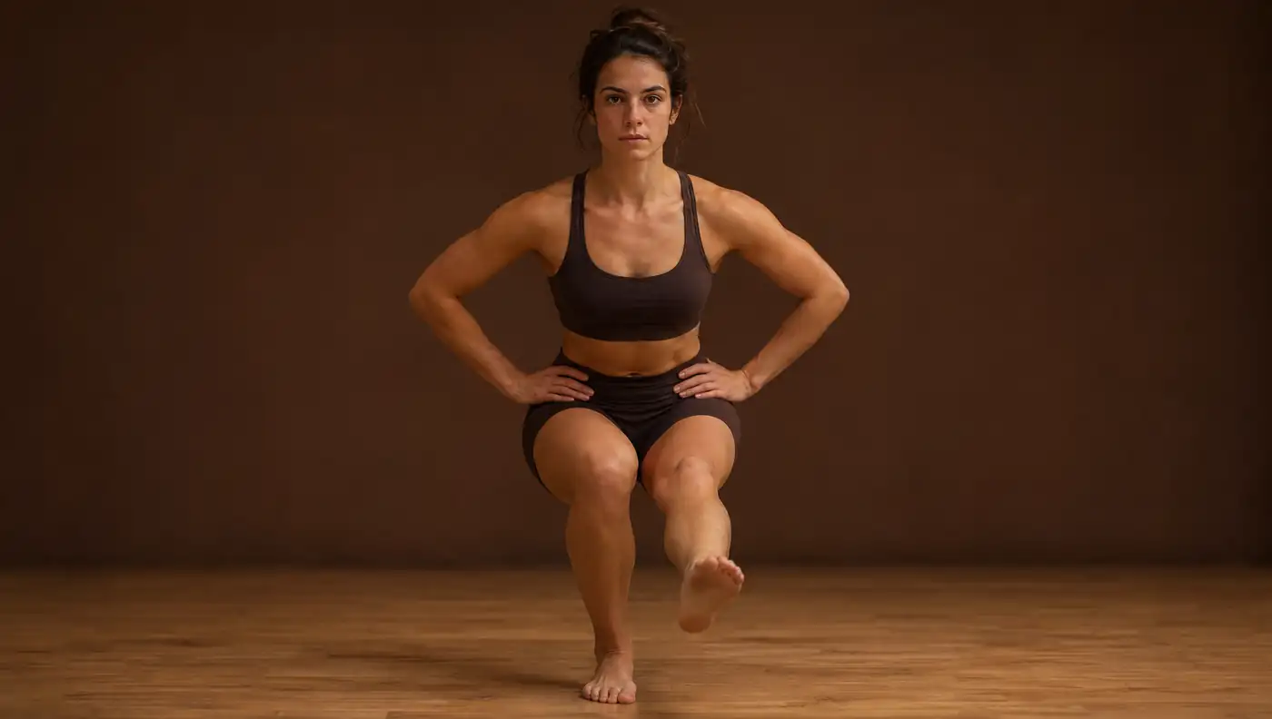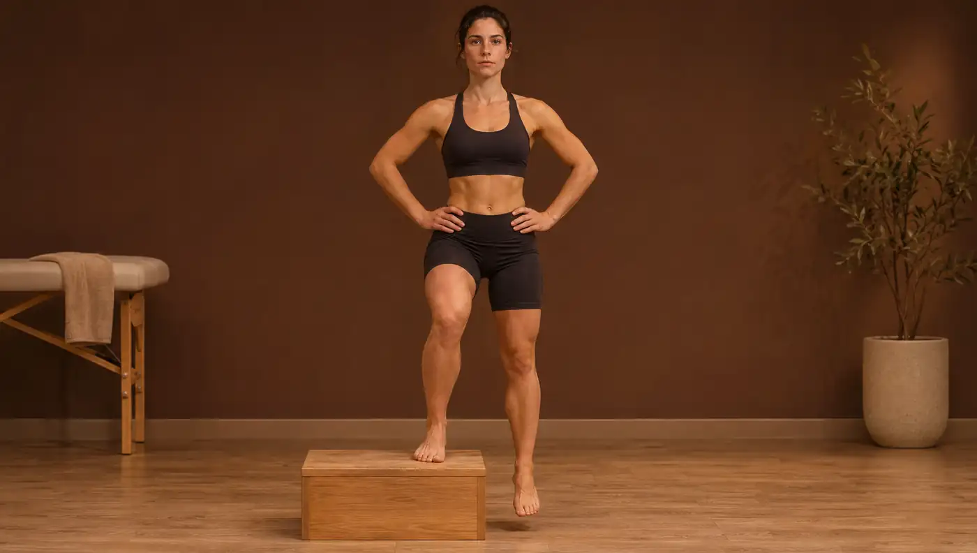
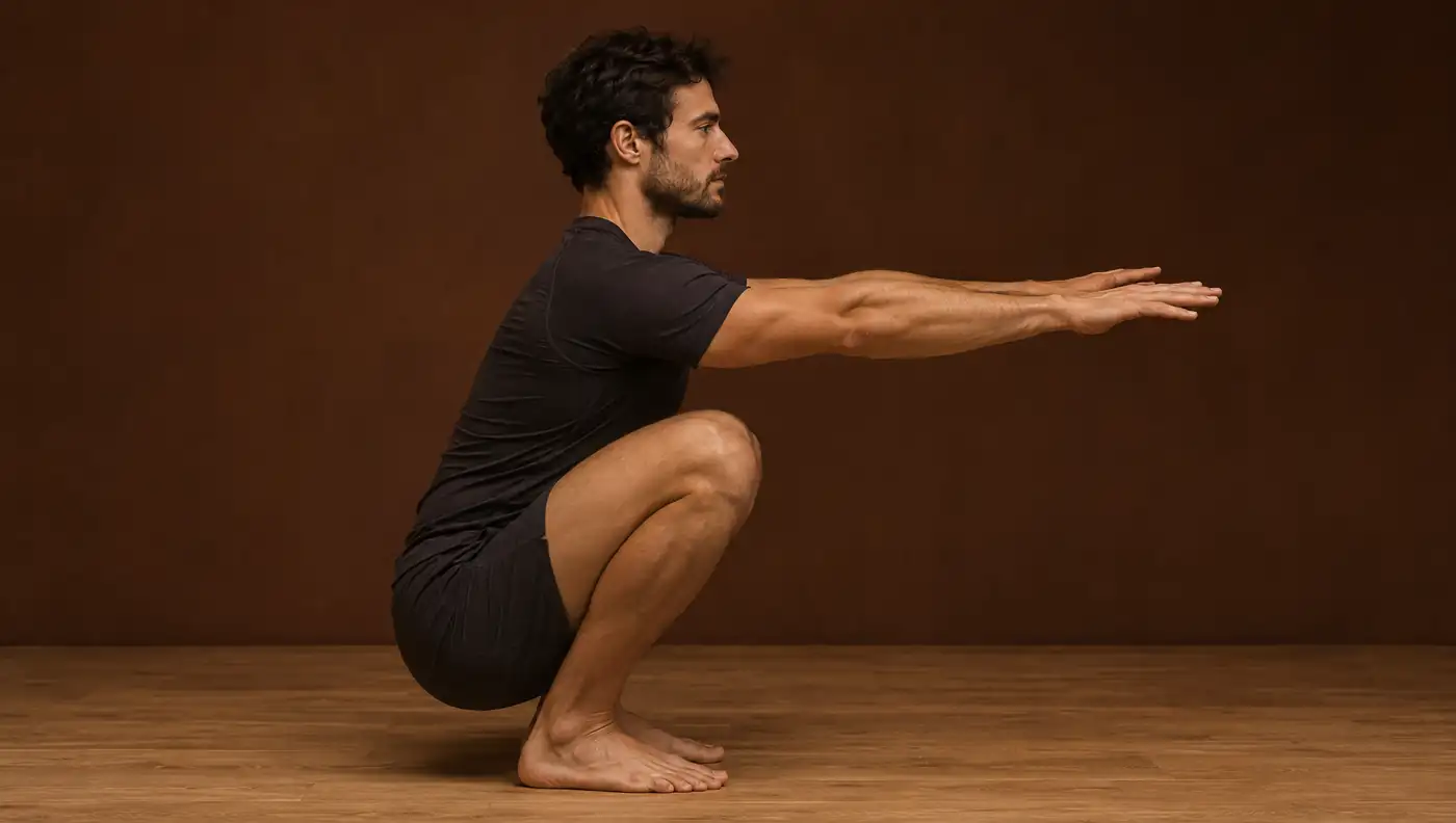
Deep squat in lateral view: ankle, knee, hip and trunk
Seen from the side, the deep squat shows how the ankle, knee, hip and trunk share the range of motion needed to descend. It is also the plane in which 2D video analysis is most valid.
- With lunge test dorsiflexion ≥ 44°, peak knee flexion in the overhead squat was 112.7 ± 13.5°, compared with 97.5 ± 14.3° with ≤ 43° 1.
- Restricting dorsiflexion with a 12° wedge reduced knee flexion and increased medial knee displacement (effect size 2.92) 2.
- Preventing forward knee travel raised hip torque from 28.2 to 302.7 N·m and increased forward trunk lean 3.
- The lunge test is highly reliable (ICC 0.80–0.99); a real change requires about 4.6–4.7° or 1.6–1.9 cm 4.
- In lateral view, 2D angles agree well with 3D (r = 0.51–0.93) 5; the hip is the least accurate angle with pose estimation 6.
Why this test matters
The bilateral deep squat is a fundamental movement pattern, found in strength training, rehabilitation and many activities of daily living. In lateral view, it shows each joint’s contribution to depth: forward travel of the tibia over the foot (ankle dorsiflexion), knee and hip flexion, and trunk lean.
The range required is large. In a kinematic study of 30 healthy adults from a population whose daily activities include squatting, the full squat with heels raised required 157 ± 6° of knee flexion, and the squat with heels down up to 95 ± 27° of hip flexion, a standard deviation that shows how much it varies from person to person 7. When one joint does not contribute, another compensates, and it is this redistribution that matters to the clinician.
The ankle as a limiting factor
Ankle dorsiflexion is the most studied determinant of squat depth. In 101 healthy adults, dorsiflexion with the knee flexed and hip flexion range explained an important part of depth in men (R² = 0.435); in women, dorsiflexion with the knee extended and dorsiflexor strength explained R² = 0.324 8.
How it is measured matters. In 40 active adults, classifying participants by weight-bearing dorsiflexion (lunge test) distinguished their squat pattern, whereas non-weight-bearing passive dorsiflexion showed no differences. The group with normal dorsiflexion (≥ 44°; mean 50.8 ± 5.2°) reached greater knee flexion in the overhead squat than the restricted group (≤ 43°; 38.9 ± 3.5°): 112.7 ± 13.5° vs 97.5 ± 14.3° 1.
Under experimental conditions, the relationship is direct: placing the feet on a 12° wedge under the forefoot, simulating a lack of plantar flexor flexibility, reduced peak knee flexion (effect size 0.81) and increased valgus (0.21) and medial knee displacement (2.92), without altering hip kinematics 2. A meta-analysis of 17 studies confirmed the association between reduced dorsiflexion and dynamic valgus (SMD −0.65), which was stronger when dorsiflexion was measured weight-bearing (SMD −1.25) 9.
Knee, hip and trunk: redistributing the load
Stopping the knee from passing the toes does not make the squat safer: it shifts the load. In seven trained men, squatting with a barrier in front of the knees reduced knee torque from 150.1 ± 50.8 to 117.3 ± 34.2 N·m, but increased hip torque from 28.2 ± 65.0 to 302.7 ± 71.2 N·m, with greater forward lean of the trunk and shank 3. Some forward tibial travel is needed to distribute load between the knee, hip and lumbar spine.
In a study of nine men analysed on 2D video, dorsiflexion range predicted the ankle angle in the squat and hip flexion on the opposite side, and knee flexion range predicted forward trunk lean 11. The sample is small, but it illustrates the clinical reasoning: increased trunk lean may be compensation for a more distal restriction rather than a trunk problem.
Standardised protocol and video capture
| Element | Published description | Source |
|---|---|---|
| Overhead variant | Feet shoulder-width apart, arms overhead, heels on the floor, at least 60° of knee flexion | 1 |
| Cadence | Metronome at 60 bpm: 2 beats down, 2 up | 1 |
| Maximum-depth variant | Hands behind the head, feet pelvis-width apart, descend as far as possible without lifting the heels, hold 5 s; 3 trials | 8 |
| Camera | Lateral, 2.4 m from the sagittal plane, at knee height, level and perpendicular to the floor | 8 |
| Depth | Vertical hip–floor distance normalised to lower-limb length | 8 |
| Trunk lean | C7–L5 line relative to the vertical | 11 |
| Knee flexion | Greater trochanter–lateral epicondyle line vs fibular head–lateral malleolus line | 11 |
The chosen variant (overhead or maximum depth) and foot position should be kept the same at every reassessment and recorded.
Reference values
| Measure | Value | Population | Source |
|---|---|---|---|
| Peak knee flexion, overhead squat (lunge ≥ 44°) | 112.7 ± 13.5° | 40 active adults | 1 |
| Peak knee flexion, overhead squat (lunge ≤ 43°) | 97.5 ± 14.3° | 40 active adults | 1 |
| Lunge test, normal vs restricted group | 50.8 ± 5.2° vs 38.9 ± 3.5° | 40 active adults | 1 |
| Peak knee flexion, heels raised | 157 ± 6° | 30 healthy adults (48.2 ± 7.6 years) | 7 |
| Peak hip flexion, heels down | 95 ± 27° | 30 healthy adults (48.2 ± 7.6 years) | 7 |
The 44° cut-off corresponds to the median of the Dill et al. sample, not to a validated norm.
There are no validated 2D normative values for depth or trunk angle in the deep squat. The values above come from 3D measurements and specific populations, so they serve as a frame of reference rather than a threshold. For individual monitoring, the most useful reference is the patient themselves, the contralateral side and dorsiflexion measured with the lunge test.
Reliability and measurement error
The sagittal plane is where 2D video is most valid. In the single-leg squat, sagittal joint displacements in 2D correlated moderately to strongly with 3D (r = 0.51–0.93), with mean differences of 0.74–3.12° 5; with pose estimation, sagittal angles at peak knee flexion correlated with 3D at r = 0.68–0.94 12. In the bilateral squat filmed from the side, OpenPose had almost perfect test–retest reliability and almost perfect or substantial agreement with the VICON system for the trunk, knee and ankle, but only fair agreement for the hip, with fixed biases at the knee and ankle and proportional biases at the trunk and hip 6.
The lunge test is the natural complement. Reliability was excellent in the original study (intra-rater ICC 0.97–0.98; inter-rater 0.97 for angle and 0.99 for distance) 13. A systematic review of 12 studies found an inter-clinician ICC of 0.80–0.99 and an intra-clinician ICC of 0.65–0.99 4. Even an inexperienced rater obtained an ICC of 0.96–0.99 with a tape measure and inclinometer 14.
Clinical interpretation and limitations
- The mechanistic studies are cross-sectional and often small: seven participants in Fry et al. 3 and nine in Endo et al. 11.
- ‘Depth’ is defined in different ways: normalised hip–floor distance 8, peak knee flexion or reaching 60° of flexion 1.
- The fixed and proportional biases of pose estimation 6 prevent values obtained from video being compared directly with marker-based 3D norms.
- If the squat is also observed from the front, valgus measured in 2D is a projection: in the single-leg squat, the pooled correlation with 3D knee abduction was only r = 0.13 15.
- No single movement test has shown good ability to predict individual injuries; in the single-leg squat, the most studied test, discrimination was poor (AUC 0.59) 16. In the studies reviewed, squat depth was not related to injury risk.
The clinical value of the test lies in identifying the joint that limits the movement and in monitoring the same person over time, together with the lunge test.
Frequently asked questions
How much ankle dorsiflexion do you need to squat?
There is no single validated value. In one study, people with ≥ 44° on the lunge test reached about 15° more knee flexion in the overhead squat than those with ≤ 43° 1.
What is a normal knee-to-wall lunge test result?
The literature reviewed does not establish a validated normative value. In active adults, the normal dorsiflexion group had 50.8 ± 5.2° and the restricted group 38.9 ± 3.5° 1; it is more useful to compare sides and track changes greater than the MDC of about 4.6–4.7° 4.
Is it dangerous to let your knees go past your toes when squatting?
Preventing forward knee travel reduced knee torque, but raised hip torque from 28.2 to 302.7 N·m and increased trunk lean 3. Some forward tibial travel is normal and necessary.
Why do my knees cave in when I squat?
One cause is restricted dorsiflexion: limiting it experimentally increased medial knee displacement 2, and a meta-analysis confirms the association (SMD −0.65) 9.
Is 2D video analysis of the squat reliable?
In lateral view, yes: pose estimation had almost perfect reliability and good agreement with 3D for the trunk, knee and ankle, but only fair agreement for the hip 6.
References
- Dill KE, Begalle RL, Frank BS, Zinder SM, Padua DA. Altered knee and ankle kinematics during squatting in those with limited weight-bearing-lunge ankle-dorsiflexion range of motion. J Athl Train. 2014;49(6):723–732. doi.org/10.4085/1062-6050-49.3.29
- Macrum E, Bell DR, Boling M, Lewek M, Padua D. Effect of limiting ankle-dorsiflexion range of motion on lower extremity kinematics and muscle-activation patterns during a squat. J Sport Rehabil. 2012;21(2):144–150. doi.org/10.1123/jsr.21.2.144
- Fry AC, Smith JC, Schilling BK. Effect of knee position on hip and knee torques during the barbell squat. J Strength Cond Res. 2003;17(4):629–633. doi.org/10.1519/1533-4287(2003)017%3C0629:EOKPOH%3E2.0.CO;2
- Powden CJ, Hoch JM, Hoch MC. Reliability and minimal detectable change of the weight-bearing lunge test: a systematic review. Man Ther. 2015;20(4):524–532. doi.org/10.1016/j.math.2015.01.004
- Schurr SA, Marshall AN, Resch JE, Saliba SA. Two-dimensional video analysis is comparable to 3D motion capture in lower extremity movement assessment. Int J Sports Phys Ther. 2017;12(2):163–172. pmc.ncbi.nlm.nih.gov/articles/PMC5380858/
- Ota M, Tateuchi H, Hashiguchi T, Kato T, Ogino Y, Yamagata M, et al. Verification of reliability and validity of motion analysis systems during bilateral squat using human pose tracking algorithm. Gait Posture. 2020;80:62–67. doi.org/10.1016/j.gaitpost.2020.05.027
- Hemmerich A, Brown H, Smith S, Marthandam SS, Wyss UP. Hip, knee, and ankle kinematics of high range of motion activities of daily living. J Orthop Res. 2006;24(4):770–781. doi.org/10.1002/jor.20114
- Kim SH, Kwon OY, Park KN, Jeon IC, Weon JH. Lower extremity strength and the range of motion in relation to squat depth. J Hum Kinet. 2015;45:59–69. doi.org/10.1515/hukin-2015-0007
- Lima YL, Ferreira VMLM, de Paula Lima PO, Bezerra MA, de Oliveira RR, Almeida GPL. The association of ankle dorsiflexion and dynamic knee valgus: a systematic review and meta-analysis. Phys Ther Sport. 2018;29:61–69. doi.org/10.1016/j.ptsp.2017.07.003
- Bell DR, Padua DA, Clark MA. Muscle strength and flexibility characteristics of people displaying excessive medial knee displacement. Arch Phys Med Rehabil. 2008;89(7):1323–1328. doi.org/10.1016/j.apmr.2007.11.048
- Endo Y, Miura M, Sakamoto M. The relationship between the deep squat movement and the hip, knee and ankle range of motion and muscle strength. J Phys Ther Sci. 2020;32(6):391–394. doi.org/10.1589/jpts.32.391
- Haberkamp LD, Garcia MC, Bazett-Jones DM. Validity of an artificial intelligence, human pose estimation model for measuring single-leg squat kinematics. J Biomech. 2022;144:111333. doi.org/10.1016/j.jbiomech.2022.111333
- Bennell KL, Talbot RC, Wajswelner H, Techovanich W, Kelly DH, Hall AJ. Intra-rater and inter-rater reliability of a weight-bearing lunge measure of ankle dorsiflexion. Aust J Physiother. 1998;44(3):175–180. doi.org/10.1016/s0004-9514(14)60377-9
- Konor MM, Morton S, Eckerson JM, Grindstaff TL. Reliability of three measures of ankle dorsiflexion range of motion. Int J Sports Phys Ther. 2012;7(3):279–287. pmc.ncbi.nlm.nih.gov/articles/PMC3362988/
- Lopes TJA, Ferrari D, Ioannidis J, Simic M, Mícolis de Azevedo F, Pappas E. Reliability and validity of frontal plane kinematics of the trunk and lower extremity measured with 2-dimensional cameras during athletic tasks: a systematic review with meta-analysis. J Orthop Sports Phys Ther. 2018;48(10):812–822. doi.org/10.2519/jospt.2018.8006
- Räisänen AM, Pasanen K, Krosshaug T, Vasankari T, Kannus P, Heinonen A, et al. Association between frontal plane knee control and lower extremity injuries: a prospective study on young team sport athletes. BMJ Open Sport Exerc Med. 2018;4(1):e000311. doi.org/10.1136/bmjsem-2017-000311
Educational content for health professionals. It does not replace clinical assessment or reading the original studies.