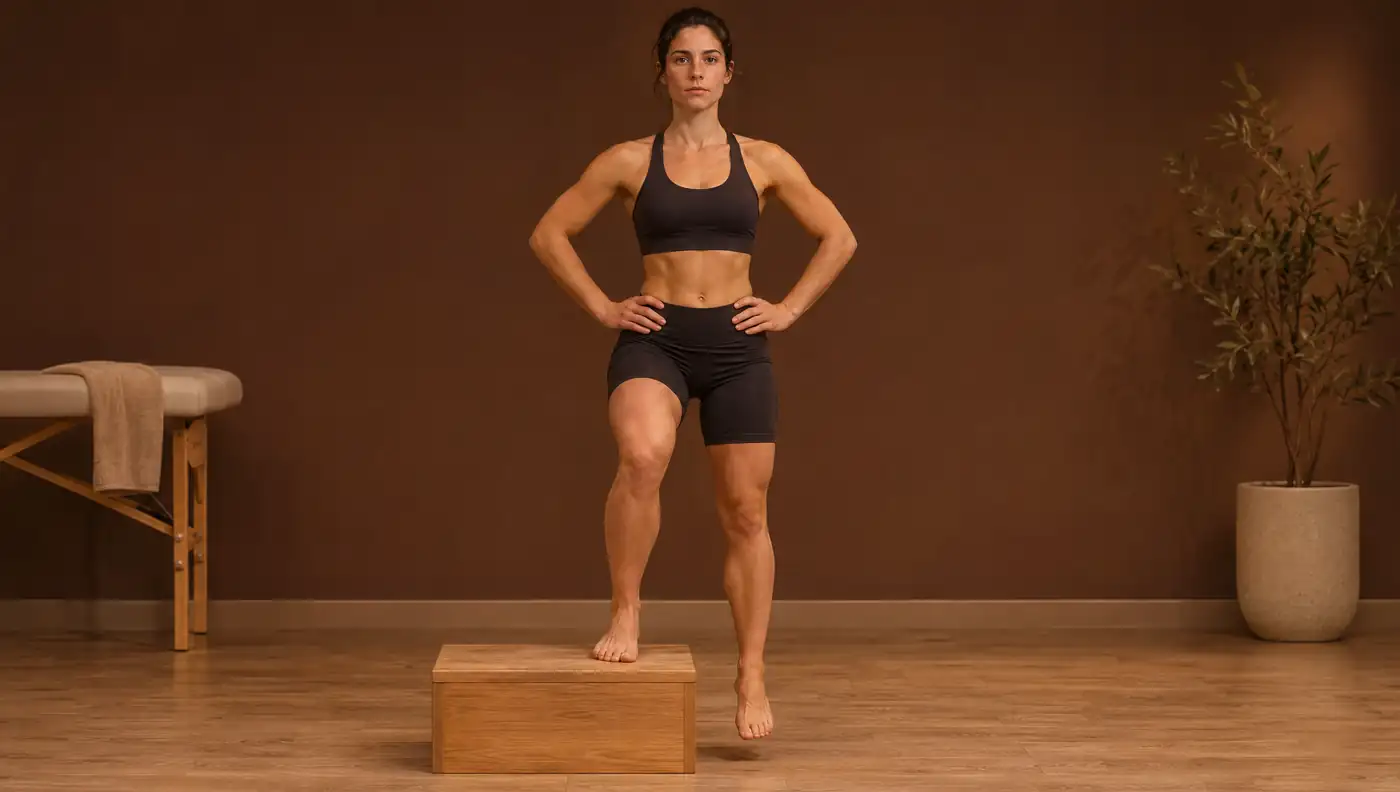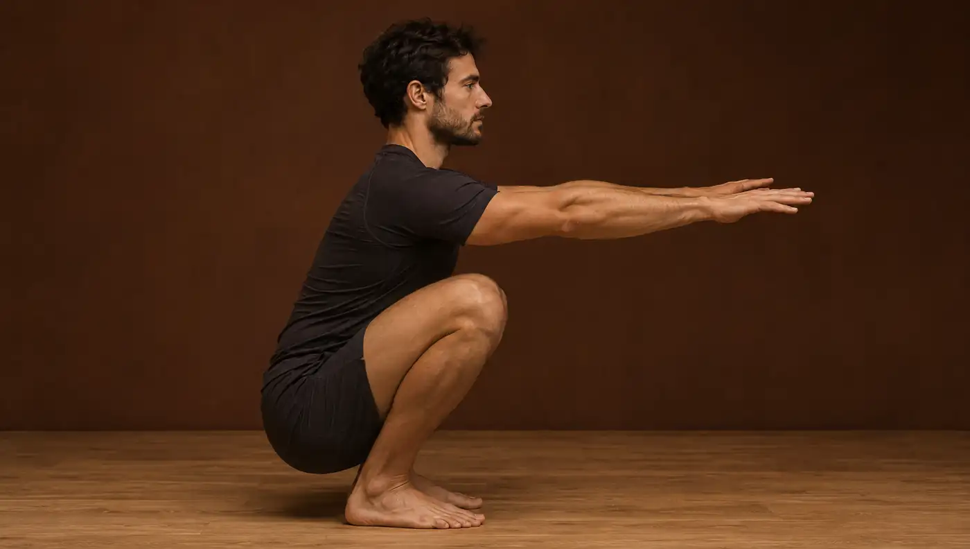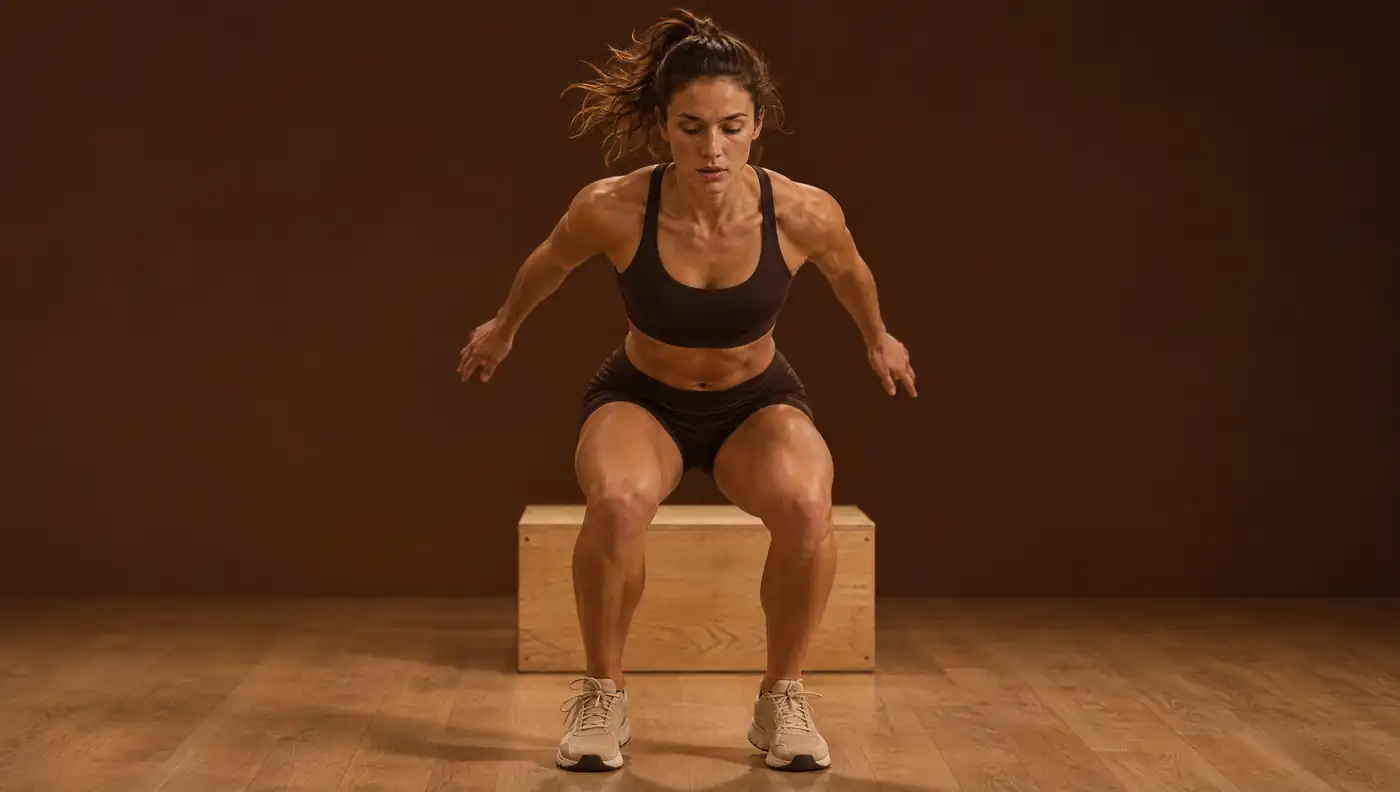
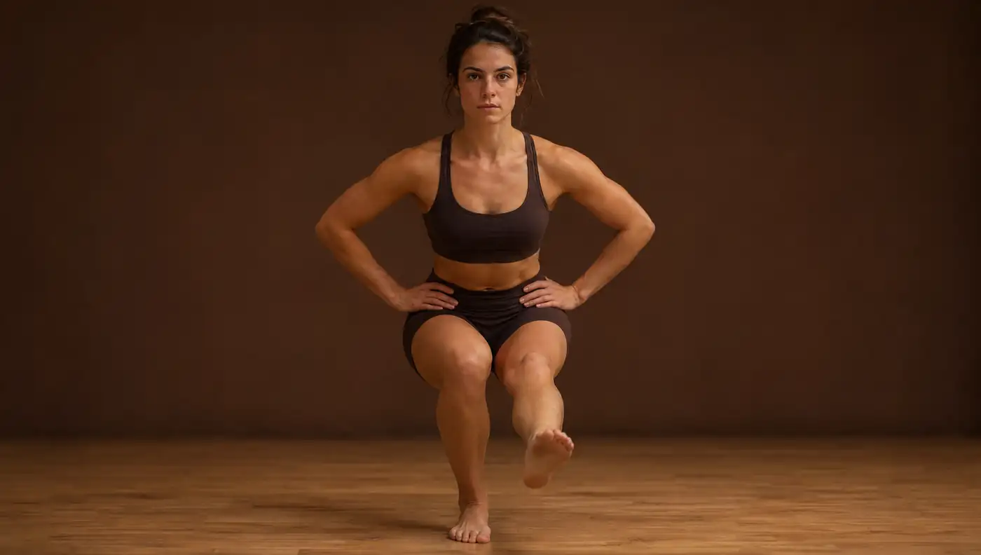
Single-leg squat: what to measure, how to film it and how to interpret it
The single-leg squat is one of the most widely used control tests in the clinic. Measured on 2D video it is reproducible and informative, provided you know what the frontal plane projection angle (FPPA) represents and what it does not.
- In asymptomatic women, mean FPPA in the single-leg squat was 8.4 ± 5.1°, compared with 16.8 ± 5.4° in women with patellofemoral pain 1.
- FPPA is a 2D projection: in the single-leg squat, the pooled correlation with knee abduction measured in 3D was only r = 0.13 2.
- Between-day reliability is good (ICC 0.72–0.91), but the smallest detectable difference is 7.5–8.9° 3.
- Trunk and pelvis add information: in patellofemoral pain there is more ipsilateral trunk lean, contralateral pelvic drop and hip adduction 4.
- As a stand-alone screening test, the single-leg squat discriminates poorly between those who will and will not get injured (AUC 0.59) 5.
Why this test matters
The single-leg squat assesses the ability to control the lower limb, pelvis and trunk under load on one leg. A knee that moves inwards, so-called dynamic knee valgus, is associated with patellofemoral pain and is often used when screening athletes and monitoring rehabilitation 1,6.
The test reflects proximal muscle function. In a study of 34 asymptomatic adults, those rated as good performers by a panel of clinicians had earlier gluteus medius activation and greater hip abduction torque than poor performers; rater agreement with the panel was 73–87% (κ = 0.600–0.800) 7. The ankle also plays a part: participants with medial knee displacement showed less passive dorsiflexion and lower gluteal coactivation relative to the hip adductors 8.
In pre-season screening, 51% of 142 young athletes had a positive single-leg squat, and they showed more dynamic valgus in a drop jump analysed in 2D 9. In 186 elite handball players, 2D analysis and a physiotherapist’s visual assessment agreed well in the single-leg squat (AUC 0.83–0.89) 10.
What we measure and how each variable is defined
FPPA is the angle, in the image plane, between the thigh line (anterior superior iliac spine to the centre of the knee) and the shank line (centre of the knee to the centre of the ankle); 0° is neutral alignment and positive values represent valgus 5. It is usually read at the frame of peak knee flexion 11.
FPPA is not true knee abduction. It is the 2D projection of a three-dimensional movement, influenced by hip rotation, knee flexion and camera orientation. In 25 adults, those whose knee moved medial to the foot showed more valgus in 2D (11.6° vs 5.0°), but not more valgus in 3D (6.1° vs 5.0°); the real difference lay in hip internal rotation (10.6° vs 4.8°) 12. In women with patellofemoral pain, FPPA was associated with hip adduction (r = 0.32–0.38) and knee external rotation (r = 0.48–0.55), and the authors caution that it should not be used to quantify 3D rotations 13.
- Hip adduction: in 2D, it correlated with adduction measured in 3D (r = 0.81) during the single-leg squat 11.
- Contralateral pelvic drop: lowering of the hemipelvis on the non-stance side; greater in patellofemoral pain 4.
- Lateral trunk lean: measured in 2D with an ICC of 0.98–1.00; in a single-leg jump task, only the sum of valgus and trunk lean was associated with the 3D knee abduction moment 14.
- Depth: the knee flexion reached; it affects all the other values and should be recorded 15.
Standardised protocol and video capture
There is no single protocol. A systematic review found wide variation in how the single-leg squat is performed, which prevented the identification of common biomechanical parameters that distinguish pathological groups, and called for standardisation 15. The most useful published variants are these:
| Parameter | Option described | Source |
|---|---|---|
| Depth | 45–60° of knee flexion | 15 |
| Depth | 60° of knee flexion | 4,6 |
| Depth | ≈50° (mini-squat: bend until the toes are no longer visible) | 12 |
| Depth | 90° of knee flexion (too demanding for many children) | 5,16 |
| Cadence | 20 squats/min with a metronome | 12 |
| Cadence | Full squat in 5 s | 11 |
| Arms | Hands on hips | 5 |
| Repetitions | 3 per limb; average of the valid ones | 5 |
The 45–60° depth corresponds to Herrington’s protocol, as described in the review by Warner et al.
The camera should face the subject, perpendicular to the frontal plane; in one validation protocol it was placed 60 cm high 11. Camera tilt or body rotation alters the projection, so foot position, camera distance and camera height should be repeated between sessions. A repetition is invalid when the free limb touches the floor, the hands leave the hips or the subject loses balance 5.
Reference values
| Population | Task | FPPA (mean ± SD) | Source |
|---|---|---|---|
| Asymptomatic women (n = 30) | Single-leg squat | 8.4 ± 5.1° | 1 |
| Women with patellofemoral pain (n = 12) | Single-leg squat | 16.8 ± 5.4° | 1 |
| Adolescent basketball and floorball athletes (n = 306) | Single-leg squat to 90° | 13.3 ± 10.5° | 5 |
| Older girls, youth football (U11–U14) | Single-leg squat to 90° | 12.2 ± 8.3° | 16 |
| Physically active adults (n = 100) | Single-leg landing from a step | Women 5–12°; men 1–9° | 17 |
The landing values are not from the single-leg squat; they serve only as a sex-specific frame of reference and for assessing symmetry.
For the other segments, the 3D values from Nakagawa et al. give an order of magnitude for patellofemoral pain compared with controls: trunk lean 9.3 ± 5.3° vs 6.7 ± 3.0°, contralateral pelvic drop 10.3 ± 4.7° vs 7.4 ± 3.8° and hip adduction 14.8 ± 7.8° vs 10.8 ± 5.6° 4. These differences of 3–4° are close to the measurement error in 2D.
The threshold of 23.8°, 1 standard deviation above the mean of an adolescent cohort, was associated with a higher risk of lower-limb injury (adjusted OR 2.67) and ankle injury (OR 2.37), but not knee injury (OR 1.49, not statistically significant) 5. It is a proposal derived from a single cohort: it should be read as ‘high relative to peers’, not as a pass/fail criterion.
Reliability and measurement error
Measured on video, FPPA is reproducible. In 20 students, within-day ICC ranged from 0.59 to 0.88 and between-day ICC from 0.72 to 0.91, with a standard error of measurement (SEM) of 2.72–3.01° and a smallest detectable difference of 7.54–8.93° 3. Other studies found an ICC of 0.86 within the same session and 0.74 between sessions, with an SEM of 2–4° 18, and an inter-rater ICC of 0.97–0.99 11.
Visual rating, without measurement, is less consistent: in a meta-analysis of 31 studies, pooled inter-rater agreement was 0.58 and intra-rater agreement 0.68 19. Automated pose estimation reproduces manual 2D measurement well: r = 0.95 between the two techniques in the single-leg squat 20, and concordance (CCC) of 0.95 with OpenPose and 0.89 with BlazePose against manual measurement 21. This means that pose estimation inherits both the strengths and the limitations of classic 2D analysis.
Clinical interpretation and limitations
- Validity against 3D. In the single-leg squat, the relationship between FPPA and 3D knee abduction ranges from absent (pooled r of 0.13 in a meta-analysis 2; r = 0.308 22; r = 0.20 with pose estimation 20) to good (r = 0.64–0.78 18; r = 0.79 11), depending on protocol and sample. Sagittal 2D measures are considerably more valid (r = 0.51–0.93) 22.
- Injury prediction. In adolescents, high FPPA discriminated poorly between future injured and uninjured athletes (AUC 0.59) 5; in 558 young footballers it was not a risk factor, and neither was side-to-side asymmetry 16. The test should not be used on its own to predict injury.
- Subgroups, not diagnosis. FPPA distinguished patients with patellofemoral pain from controls (effect size 0.68), with only fair sensitivity and specificity 6. It helps to identify who presents with dynamic valgus.
- More than one test. The single-leg squat and the drop jump identified different players with poor knee control 10.
A systematic review concluded that FPPA has acceptable reliability and construct validity, but unacceptable criterion validity: it does not replace 3D analysis 23. In practice, it is an index of movement quality, useful for describing the pattern, comparing sides and following the same patient with the same protocol, always as part of the clinical assessment.
Frequently asked questions
What is a normal FPPA in the single-leg squat?
In asymptomatic women the mean was 8.4 ± 5.1°; in women with patellofemoral pain, 16.8 ± 5.4° 1. Values depend on depth and protocol, so there is no universal ‘normal’.
Does FPPA measure true knee valgus?
No. It is a 2D projection that, in the single-leg squat, correlates weakly with 3D knee abduction (pooled r of 0.13) 2 and mainly reflects hip adduction and internal rotation 12.
How many degrees of change in FPPA are clinically meaningful?
No MCID has been published. The between-day smallest detectable difference is 7.5–8.9° 3; smaller changes may be measurement error.
Does the single-leg squat predict knee injuries?
Not in a way that is useful for an individual. In an adolescent cohort, an FPPA above 23.8° was not associated with knee injury, and overall discriminative ability was poor (AUC 0.59) 5.
References
- Herrington L. Knee valgus angle during single leg squat and landing in patellofemoral pain patients and controls. Knee. 2014;21(2):514–517. doi.org/10.1016/j.knee.2013.11.011
- Lopes TJA, Ferrari D, Ioannidis J, Simic M, Mícolis de Azevedo F, Pappas E. Reliability and validity of frontal plane kinematics of the trunk and lower extremity measured with 2-dimensional cameras during athletic tasks: a systematic review with meta-analysis. J Orthop Sports Phys Ther. 2018;48(10):812–822. doi.org/10.2519/jospt.2018.8006
- Munro A, Herrington L, Carolan M. Reliability of 2-dimensional video assessment of frontal-plane dynamic knee valgus during common athletic screening tasks. J Sport Rehabil. 2012;21(1):7–11. doi.org/10.1123/jsr.21.1.7
- Nakagawa TH, Moriya ÉT, Maciel CD, Serrão FV. Trunk, pelvis, hip, and knee kinematics, hip strength, and gluteal muscle activation during a single-leg squat in males and females with and without patellofemoral pain syndrome. J Orthop Sports Phys Ther. 2012;42(6):491–501. doi.org/10.2519/jospt.2012.3987
- Räisänen AM, Pasanen K, Krosshaug T, Vasankari T, Kannus P, Heinonen A, et al. Association between frontal plane knee control and lower extremity injuries: a prospective study on young team sport athletes. BMJ Open Sport Exerc Med. 2018;4(1):e000311. doi.org/10.1136/bmjsem-2017-000311
- Gwynne CR, Curran SA. Two-dimensional frontal plane projection angle can identify subgroups of patellofemoral pain patients who demonstrate dynamic knee valgus. Clin Biomech (Bristol). 2018;58:44–48. doi.org/10.1016/j.clinbiomech.2018.06.021
- Crossley KM, Zhang WJ, Schache AG, Bryant A, Cowan SM. Performance on the single-leg squat task indicates hip abductor muscle function. Am J Sports Med. 2011;39(4):866–873. doi.org/10.1177/0363546510395456
- Mauntel TC, Begalle RL, Cram TR, Frank BS, Hirth CJ, Blackburn T, et al. The effects of lower extremity muscle activation and passive range of motion on single leg squat performance. J Strength Cond Res. 2013;27(7):1813–1823. doi.org/10.1519/JSC.0b013e318276b886
- Ugalde V, Brockman C, Bailowitz Z, Pollard CD. Single leg squat test and its relationship to dynamic knee valgus and injury risk screening. PM R. 2015;7(3):229–235. doi.org/10.1016/j.pmrj.2014.08.361
- Stensrud S, Myklebust G, Kristianslund E, Bahr R, Krosshaug T. Correlation between two-dimensional video analysis and subjective assessment in evaluating knee control among elite female team handball players. Br J Sports Med. 2011;45(7):589–595. doi.org/10.1136/bjsm.2010.078287
- Herrington L, Alenezi F, Alzhrani M, Alrayani H, Jones R. The reliability and criterion validity of 2D video assessment of single leg squat and hop landing. J Electromyogr Kinesiol. 2017;34:80–85. doi.org/10.1016/j.jelekin.2017.04.004
- Ageberg E, Bennell KL, Hunt MA, Simic M, Roos EM, Creaby MW. Validity and inter-rater reliability of medio-lateral knee motion observed during a single-limb mini squat. BMC Musculoskelet Disord. 2010;11:265. doi.org/10.1186/1471-2474-11-265
- Willson JD, Davis IS. Utility of the frontal plane projection angle in females with patellofemoral pain. J Orthop Sports Phys Ther. 2008;38(10):606–615. doi.org/10.2519/jospt.2008.2706
- Dingenen B, Malfait B, Vanrenterghem J, Verschueren SM, Staes FF. The reliability and validity of the measurement of lateral trunk motion in two-dimensional video analysis during unipodal functional screening tests in elite female athletes. Phys Ther Sport. 2014;15(2):117–123. doi.org/10.1016/j.ptsp.2013.05.001
- Warner MB, Wilson DA, Herrington L, Dixon S, Power C, Jones R, et al. A systematic review of the discriminating biomechanical parameters during the single leg squat. Phys Ther Sport. 2019;36:78–91. doi.org/10.1016/j.ptsp.2019.01.007
- Räisänen AM, Arkkila H, Vasankari T, Steffen K, Parkkari J, Kannus P, et al. Investigation of knee control as a lower extremity injury risk factor: a prospective study in youth football. Scand J Med Sci Sports. 2018;28(9):2084–2092. doi.org/10.1111/sms.13197
- Herrington L, Munro A. Drop jump landing knee valgus angle; normative data in a physically active population. Phys Ther Sport. 2010;11(2):56–59. doi.org/10.1016/j.ptsp.2009.11.004
- Gwynne CR, Curran SA. Quantifying frontal plane knee motion during single limb squats: reliability and validity of 2-dimensional measures. Int J Sports Phys Ther. 2014;9(7):898–906. pmc.ncbi.nlm.nih.gov/articles/PMC4275194/
- Ressman J, Grooten WJA, Rasmussen Barr E. Visual assessment of movement quality in the single leg squat test: a review and meta-analysis of inter-rater and intrarater reliability. BMJ Open Sport Exerc Med. 2019;5(1):e000541. doi.org/10.1136/bmjsem-2019-000541
- Haberkamp LD, Garcia MC, Bazett-Jones DM. Validity of an artificial intelligence, human pose estimation model for measuring single-leg squat kinematics. J Biomech. 2022;144:111333. doi.org/10.1016/j.jbiomech.2022.111333
- Janisch L, Maack LM, Fohrmann D, Marques CJ. Assessment of dynamic knee angle deviations in the frontal plane in physiotherapy clinical practice: intra- and inter-rater reliability of an application and agreement with two AI-models. BMC Musculoskelet Disord. 2026;27(1). doi.org/10.1186/s12891-026-09958-9
- Schurr SA, Marshall AN, Resch JE, Saliba SA. Two-dimensional video analysis is comparable to 3D motion capture in lower extremity movement assessment. Int J Sports Phys Ther. 2017;12(2):163–172. pmc.ncbi.nlm.nih.gov/articles/PMC5380858/
- Lally EM, Ericksen H, Earl-Boehm J. Measurement properties of clinically accessible movement assessment tools for analyzing single-leg squats and step-downs: a systematic review. J Sport Rehabil. 2022;31(4):476–489. doi.org/10.1123/jsr.2021-0287
Educational content for health professionals. It does not replace clinical assessment or reading the original studies.