Lateral step-down: movement quality, FPPA and the role of the ankle
The lateral step-down demands eccentric control on one leg from a step. It is simple to administer, has an established scoring scale and often reveals a limitation that is not at the knee: the ankle.
- The original protocol uses a 20 cm step and 5 repetitions, scored from 0 to 6: 0–1 good, 2–3 moderate and ≥ 4 poor movement quality 1.
- Visual rating has moderate reliability (κ 0.67) 1 and was only fair to moderate when done from video (κ 0.40–0.65) 2.
- FPPA measured on 2D video is reproducible (ICC 0.85), but the minimal detectable change is 9.20° 3.
- Restricted dorsiflexion is the most consistent finding in people who perform the test poorly: about 6° separates good from poor quality 4.
- The pelvis criterion is the most valid against 3D: it is associated with contralateral pelvic drop (effect size 1.65) 5.
Why this test matters
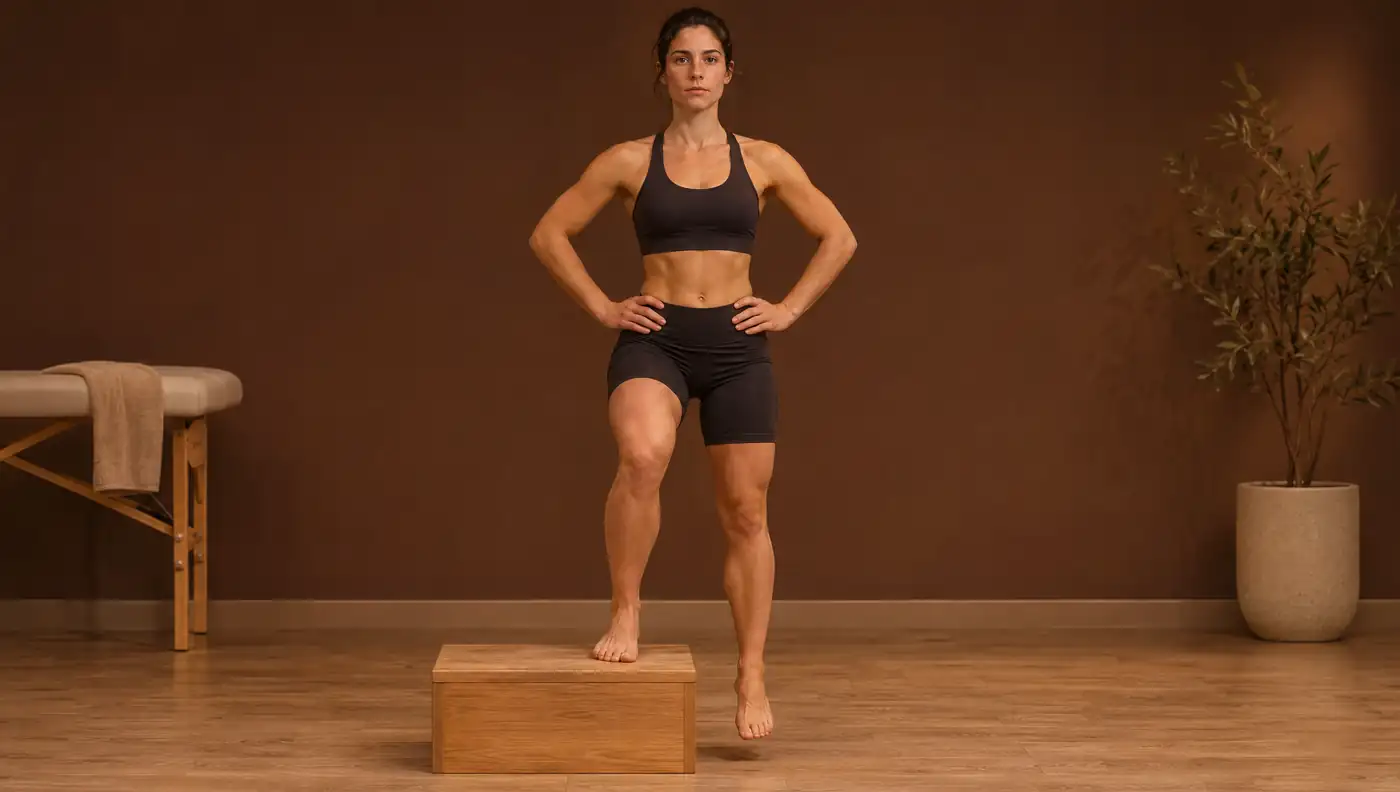
The lateral step-down test was developed to assess movement quality in patients with patellofemoral pain, based on the changes in lower-limb function commonly seen in this population 1. The patient slowly steps down from a step on one leg while the clinician observes the trunk, pelvis, knee and balance. Compared with the single-leg squat, lowering off the edge of the step places more emphasis on eccentric control.
The test is also used after anterior cruciate ligament (ACL) reconstruction: in 20 patients at least one year after surgery, the quality score from 2D video correlated with hip adduction and internal rotation measured in 3D (r = 0.47–0.57) 6. In chronic ankle instability, the test score is related to dorsiflexion 4.
What is scored and how each criterion is defined
The original scale adds points for each deviation observed during the five repetitions 1:
| Criterion | Deviation observed | Points |
|---|---|---|
| Arm strategy | Uses the arms to recover balance | 1 |
| Trunk | Leans to either side | 1 |
| Pelvis | Rotates or elevates one side relative to the other | 1 |
| Knee | Medial deviation: the tibial tuberosity passes the vertical line of the 2nd toe | 1 |
| Knee | The tibial tuberosity passes the vertical line of the medial border of the foot | 2 |
| Stance | Puts the contralateral foot down or the tested limb wobbles | 1 |
Total 0–1: good; 2–3: moderate; ≥ 4: poor movement quality 1.
Each criterion corresponds to a variable that can be measured on video. Validation against 3D showed that a pelvis rated as incorrect is associated with greater contralateral pelvic drop (effect size 1.65) and greater hip adduction (1.04); a knee rated as incorrect is associated with greater knee external rotation (0.78), and moderate overall quality with more pelvic drop (1.07) and more knee external rotation (0.72) 5.
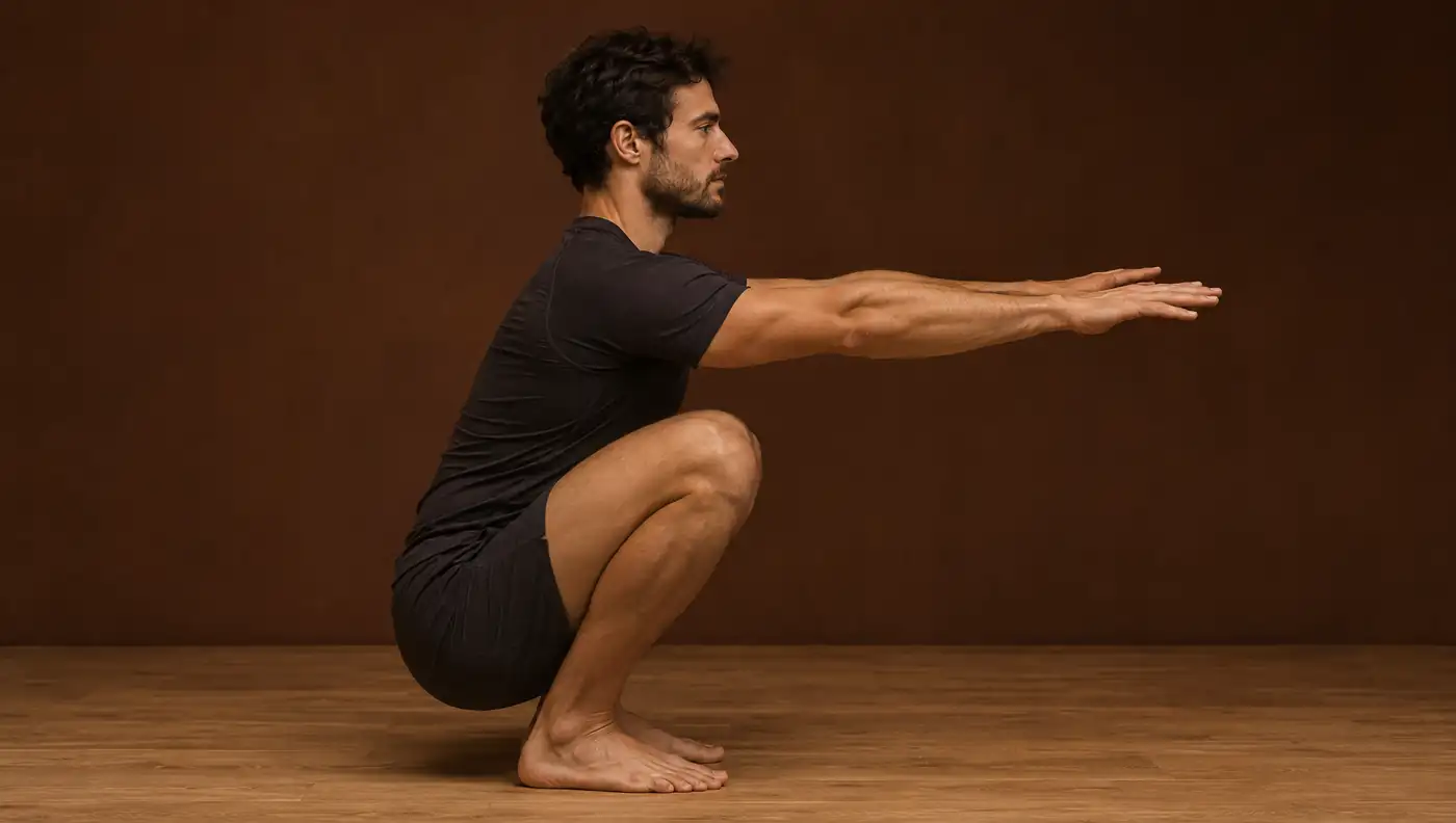
When the knee is quantified with the frontal plane projection angle (FPPA), it is worth remembering that this is a 2D projection. A systematic review concluded that FPPA is reliable in the single-leg squat and the step-down, but that its criterion validity against 3D is unacceptable 7; in the single-leg squat, the pooled correlation with 3D knee abduction was only r = 0.13 8. FPPA describes visible alignment, not true knee abduction.
Standardised protocol and video capture
- Step: 20 cm in the original protocol 1. After ACL reconstruction, 4- and 6-inch boxes (about 10 and 15 cm) were used; the higher box produced poorer movement quality 6.
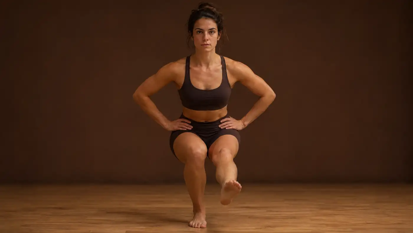
- Starting position: hands on hips, knee extended, foot close to the edge of the step; the contralateral limb is held extended over the floor beside the step 1.
- Movement: bend the tested knee until the contralateral limb lightly touches the floor, then return to extension 1.
- Repetitions: 5 per limb 1.
- Observation: the examiner stands facing the patient 1; FPPA is measured on 2D video with the patient filmed from the front 3.
What explains poor movement quality
The most consistent finding is restricted ankle dorsiflexion. In 29 healthy women, those with moderate quality had less weight-bearing dorsiflexion with the knee flexed, for both examiners, whereas hip strength did not distinguish the groups 9. In 55 male recruits, the association was repeated on the dominant side, although it was weaker than in women 10.
In 79 military personnel with patellofemoral pain, weight-bearing dorsiflexion correlated with movement quality (r = −0.39 in women; r = −0.46 in men). Among those with more than 25° of non-weight-bearing dorsiflexion, those who moved well had greater strength of the hip external rotators and knee extensors 11. In the laboratory, those with less dorsiflexion showed more hip adduction and more knee external rotation and, in the group with less weight-bearing dorsiflexion, less knee flexion 12.
| Lateral step-down quality | Weight-bearing dorsiflexion (mean ± SD) |
|---|---|
| Good | 42.3 ± 6.4° |
| Moderate | 39.1 ± 6.2° |
| Poor | 36.0 ± 6.6° |
Correlation between dorsiflexion and test score: r = −0.39 4.
In the anterior step-down, a closely related variant, lower hip abductor strength and reduced flexibility of the quadriceps and the iliotibial band/tensor fasciae latae complex distinguished women with moderate quality, with inter-rater agreement of κ = 0.80 13.
Reliability and measurement error
The visual score has moderate reliability: κ of 0.67 (80% agreement; 95% CI 0.58–0.76) between examiners in the original study of 30 patients with patellofemoral pain 1. When scoring is done from 2D video, inter- and intra-rater agreement was κ 0.40–0.65, with no difference between experienced and inexperienced physiotherapists 2. A meta-analysis that included the single-leg squat and step-down tests estimated pooled inter-rater agreement of 0.58 and intra-rater agreement of 0.68 14.
Measuring the angle, rather than rating it in categories, improves consistency: six physiotherapists measured FPPA in the lateral step-down with an ICC of 0.85 (95% CI 0.72–0.93), but with a standard error of measurement (SEM) of 3.33° and a minimal detectable change (MDC) of 9.20° 3.
Clinical interpretation and limitations
- In patellofemoral pain, lateral step-down FPPA did not correlate with pain, function (AKPS) or kinesiophobia (TSK) 3: a better angle does not necessarily mean less pain.
- The ability of the lateral step-down to predict injury has not been demonstrated. Even in the single-leg squat, the most studied control test, discrimination of future injured athletes was poor (AUC 0.59) 15.
- Most of the evidence uses visual ratings rather than angles, and step height varies between studies 6.
- Measuring weight-bearing dorsiflexion when movement quality is poor is the most consistent recommendation in the literature 9,11,4.
In practice, the lateral step-down is useful for describing the movement pattern, deciding what to assess next (ankle, hip, quadriceps) and following the same patient with a fixed protocol. The diagnostic decision remains with the clinician.
Frequently asked questions
How is the lateral step-down test scored?
Points are added for arm strategy, trunk lean, pelvic deviation, medial knee deviation (1 or 2 points) and unstable stance: 0–1 good, 2–3 moderate and ≥ 4 poor quality 1.
What step height is used for the lateral step-down test?
20 cm in the original protocol 1. Higher steps worsen the score 6, so the height should always be the same.
Why do patients with patellofemoral pain fail the lateral step-down?
Restricted weight-bearing dorsiflexion is the most strongly associated factor (r = −0.39 to −0.46); when dorsiflexion is sufficient, hip and knee extensor strength carry more weight 11.
References
- Piva SR, Fitzgerald K, Irrgang JJ, Jones S, Hando BR, Browder DA, et al. Reliability of measures of impairments associated with patellofemoral pain syndrome. BMC Musculoskelet Disord. 2006;7:33. doi.org/10.1186/1471-2474-7-33
- Mansfield C, Spech C, Rethman K, Clagg S, Ingle A, Largent A, et al. Moderate reliability of the lateral step down test amongst experienced and novice physical therapists. Physiother Theory Pract. 2022;38(12):2029–2037. doi.org/10.1080/09593985.2021.1923097
- Mansfield CJ, Rethman KK, Stephens J, Di Stasi S, Vanetten L, Briggs MS. The association and reliability of the frontal plane projection angle during the lateral step down test on knee function in patients with patellofemoral pain. Knee. 2022;36:87–96. doi.org/10.1016/j.knee.2022.04.010
- Grindstaff TL, Dolan N, Morton SK. Ankle dorsiflexion range of motion influences Lateral Step Down Test scores in individuals with chronic ankle instability. Phys Ther Sport. 2017;23:75–81. doi.org/10.1016/j.ptsp.2016.07.008
- Rabin A, Portnoy S, Kozol Z. The association between visual assessment of quality of movement and three-dimensional analysis of pelvis, hip, and knee kinematics during a lateral step down test. J Strength Cond Res. 2016;30(11):3204–3211. doi.org/10.1519/JSC.0000000000001420
- Mostaed MF, Werner DM, Barrios JA. 2D and 3D kinematics during lateral step-down testing in individuals with anterior cruciate ligament reconstruction. Int J Sports Phys Ther. 2018;13(1):77–85. pmc.ncbi.nlm.nih.gov/articles/PMC5808016/
- Lally EM, Ericksen H, Earl-Boehm J. Measurement properties of clinically accessible movement assessment tools for analyzing single-leg squats and step-downs: a systematic review. J Sport Rehabil. 2022;31(4):476–489. doi.org/10.1123/jsr.2021-0287
- Lopes TJA, Ferrari D, Ioannidis J, Simic M, Mícolis de Azevedo F, Pappas E. Reliability and validity of frontal plane kinematics of the trunk and lower extremity measured with 2-dimensional cameras during athletic tasks: a systematic review with meta-analysis. J Orthop Sports Phys Ther. 2018;48(10):812–822. doi.org/10.2519/jospt.2018.8006
- Rabin A, Kozol Z. Measures of range of motion and strength among healthy women with differing quality of lower extremity movement during the lateral step-down test. J Orthop Sports Phys Ther. 2010;40(12):792–800. doi.org/10.2519/jospt.2010.3424
- Rabin A, Kozol Z, Spitzer E, Finestone A. Ankle dorsiflexion among healthy men with different qualities of lower extremity movement. J Athl Train. 2014;49(5):617–623. doi.org/10.4085/1062-6050-49.3.14
- Rabin A, Kozol Z, Moran U, Efergan A, Geffen Y, Finestone AS. Factors associated with visually assessed quality of movement during a lateral step-down test among individuals with patellofemoral pain. J Orthop Sports Phys Ther. 2014;44(12):937–946. doi.org/10.2519/jospt.2014.5507
- Rabin A, Portnoy S, Kozol Z. The association of ankle dorsiflexion range of motion with hip and knee kinematics during the lateral step-down test. J Orthop Sports Phys Ther. 2016;46(11):1002–1009. doi.org/10.2519/jospt.2016.6621
- Park KM, Cynn HS, Choung SD. Musculoskeletal predictors of movement quality for the forward step-down test in asymptomatic women. J Orthop Sports Phys Ther. 2013;43(7):504–510. doi.org/10.2519/jospt.2013.4073
- Ressman J, Grooten WJA, Rasmussen Barr E. Visual assessment of movement quality in the single leg squat test: a review and meta-analysis of inter-rater and intrarater reliability. BMJ Open Sport Exerc Med. 2019;5(1):e000541. doi.org/10.1136/bmjsem-2019-000541
- Räisänen AM, Pasanen K, Krosshaug T, Vasankari T, Kannus P, Heinonen A, et al. Association between frontal plane knee control and lower extremity injuries: a prospective study on young team sport athletes. BMJ Open Sport Exerc Med. 2018;4(1):e000311. doi.org/10.1136/bmjsem-2017-000311
Educational content for health professionals. It does not replace clinical assessment or reading the original studies.