
Injury screening: why movement tests do not predict injuries — and what they are for
A test can be associated with injury and still be unable to predict who will get injured. Understanding this difference changes how movement tests are used and how their results are communicated.
- Validating a screening test requires three steps, and no sports injury test has met them to date 1.
- The study that launched the drop jump as an ACL screening test had 9 injuries in 205 athletes 2.
- In 710 elite athletes with 42 ruptures, the AUC for medial knee displacement was 0.6 3.
- Visual rating of the drop jump and single-leg squat had an AUC of 0.43–0.54 in 880 athletes 4.
- A systematic review found inconsistent evidence that poor movement quality increases injury risk 5.
Association is not prediction
Many studies find mean differences between athletes who get injured and those who do not. This helps to understand injury mechanisms, but it does not guarantee that the test can correctly classify an individual athlete. Because test results are continuous, there is considerable overlap between high-risk and low-risk athletes 1.
Two examples. In a cohort of young female basketball and floorball players, less knee flexion (HR 0.55 per additional 10°) and greater vertical force (HR 1.26 per 100 N) were associated with ACL rupture; even so, AUCs of 0.6 and 0.7 led the authors to conclude that these variables are not suitable for screening 6. In a Norwegian elite cohort, medial knee displacement had an OR of 1.40 per standard deviation and yet an AUC of only 0.6 3.
Statistical association answers the question ‘is this factor linked to injury in the group?’. Prediction answers a different one: ‘will this athlete, with this result, get injured?’. In clinical practice it is the second question that matters, and that is where the tests fail.
Sensitivity, specificity and AUC in plain language
- Sensitivity: the proportion of future injured athletes that the test flags.
- Specificity: the proportion of those who do not get injured that the test correctly classifies as not at risk.
- AUC: the overall ability to separate the two groups. An AUC of 0.5 is equivalent to tossing a coin; values of around 0.6–0.7 are poor to fair.
- Positive predictive value: of the athletes flagged, how many actually get injured. It depends heavily on how common the injury is in the population.
| Study | Sample and injuries | Test | Result |
|---|---|---|---|
| Hewett 2005 | 205 athletes; 9 ACL ruptures | Abduction moment in the DVJ (3D) | Sensitivity 78%, specificity 73% 2 |
| Dingenen 2015 | 50 athletes; 7 knee injuries | 2D valgus and trunk lean, single-leg DVJ | AUC 0.803 7 |
| Krosshaug 2016 | 710 athletes; 42 ruptures | Medial knee displacement, DVJ (3D) | AUC 0.6 3 |
| Leppänen 2017 | 171 athletes; 15 ruptures | Knee flexion and vertical force, DVJ (3D) | AUC 0.6–0.7 6 |
| Petushek 2021 | 880 athletes; 65 ruptures | Visual rating of the DVJ and single-leg squat | AUC 0.43–0.54 4 |
The highest AUCs appear in samples with few injuries; in the larger cohorts, predictive ability approaches chance.
From enthusiasm to replication: the case of the drop jump
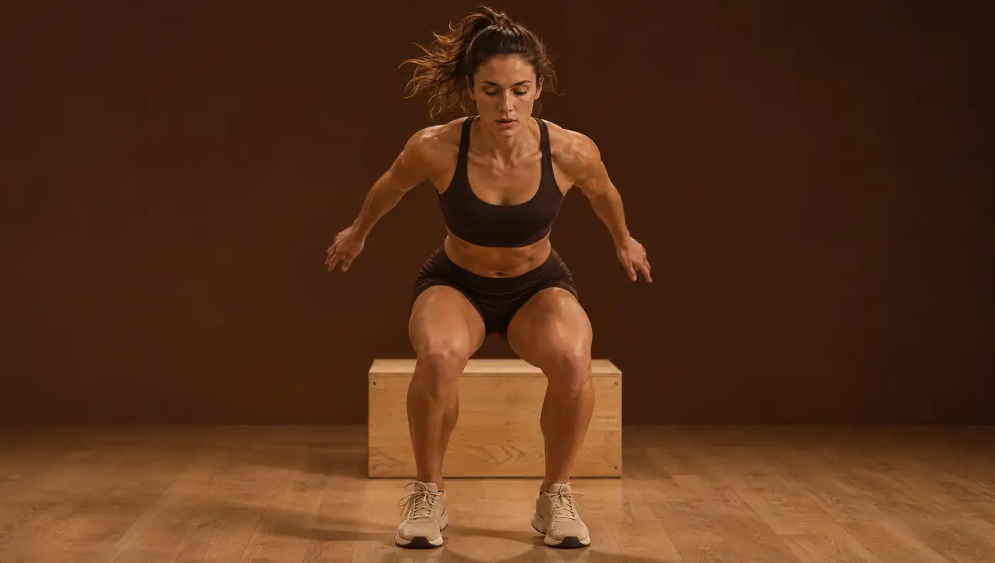
In 2005, the 9 athletes who went on to rupture their ACL landed with 8° more knee abduction and an abduction moment 2.5 times greater than the remaining 196 2. The finding launched the drop vertical jump as a screening test.
Large-scale replication did not confirm it. In the Norwegian cohort, with 782 athletes tested and 710 analysed, previous ACL injury multiplied risk (RR 3.8), but in the 643 athletes without previous injury none of the five DVJ variables was associated with risk 3. The clinical version also failed: 21% of the athletes who were later injured had a poor DVJ rating, compared with 27% of those who were not 4.
The 2D studies with positive results are small. One had 7 injuries 7. Another compared 27 ruptures with 27 controls and found peak valgus of 8.3 ± 4.3 cm versus 5.1 ± 4.1 cm, with widely overlapping standard deviations 8. A clinical algorithm estimates a high abduction moment with a sensitivity of 77% and a specificity of 71% 9, but the link between that moment and injury is precisely what the larger cohorts did not confirm 3.
What the reviews say
Bahr describes three steps needed to validate a screening test 1:
- demonstrate, in prospective studies, a strong relationship between the marker and injury risk;
- verify the test’s properties in relevant populations, using appropriate statistical tools;
- show that intervening only in flagged athletes is more beneficial than applying the same intervention to everyone.
According to the author, there is currently no sports injury screening test with adequate properties, and no intervention study that supports screening for injury risk 1.
Systematic reviews point the same way. In a review of movement quality, 17 studies were included from 4361 records, most of them low-quality cohorts (median of 11 out of 33 on the Downs and Black scale); 15 used the Functional Movement Screen, 7 reported diagnostic accuracy and none tested an intervention targeted at the higher-risk athletes 5. In another review, of ACL screening methods usable outside the laboratory, 20 studies and five methods showed limited evidence of predictive ability; the Landing Error Scoring System only showed predictive validity in a youth population 10.
So what are movement tests for?
- Profiling: describing how the athlete lands, stabilises and distributes load between sides.
- Monitoring: comparing the athlete with themselves beyond the measurement error — about 8–9° for FPPA on 2D video 11 and, calculated from the published standard error, about 0.4–0.7 s for time to stabilisation 12.
- Guiding training: normalised knee separation increased from 51% to 68% after neuromuscular training 13; time to stabilisation allows plyometric exercises to be ranked by difficulty 14.
- Deciding as part of a battery: in return to sport after ACL reconstruction, functional tests work together with strength and time since surgery. Failing to meet the six discharge criteria was associated with a 4.1 times greater risk of graft rupture 15; in another cohort, delaying return until 9 months and quadriceps strength symmetry reduced reinjury 16.
It is also worth separating what can be seen from what the knee sustains. Visual rating of valgus correlated moderately with the angle, but poorly with the abduction moment (0.09–0.11) 17. A 2D measure describes the movement pattern; it does not measure the load on the ligament.
How to communicate results to the athlete and the team
Language matters. Phrases such as ‘high risk of ACL rupture’ based on a single test are not supported by the evidence and can create anxiety or false reassurance. It is more honest to describe what was measured, compare it with a published reference value where one exists and set a date for remeasuring.
For coaches and performance staff, the argument is simple: until there is evidence that intervening only in flagged athletes is better than intervening in all of them 1, tests serve mainly to individualise progression and confirm the response to training, not to choose who receives prevention.
Frequently asked questions
Can the drop jump be used to screen for ACL injury?
Not to predict individual injuries: in 710 athletes the AUC was 0.6 3, and visual rating had an AUC of 0.43–0.54 in 880 athletes 4. It is useful for profiling and monitoring knee control.
Why don’t screening tests predict injuries?
Because results are continuous and high-risk and low-risk athletes overlap; no test has shown adequate properties, or a benefit from intervening only in flagged athletes 1.
What does an AUC of 0.6 mean in a screening test?
It means the test separates future injured and uninjured athletes only slightly better than chance, which corresponds to 0.5. This was the value found for medial knee displacement in the drop jump 3.
References
- Bahr R. Why screening tests to predict injury do not work—and probably never will…: a critical review. Br J Sports Med. 2016;50(13):776–780. doi.org/10.1136/bjsports-2016-096256
- Hewett TE, Myer GD, Ford KR, Heidt RS Jr, Colosimo AJ, McLean SG, et al. Biomechanical measures of neuromuscular control and valgus loading of the knee predict anterior cruciate ligament injury risk in female athletes: a prospective study. Am J Sports Med. 2005;33(4):492–501. doi.org/10.1177/0363546504269591
- Krosshaug T, Steffen K, Kristianslund E, Nilstad A, Mok KM, Myklebust G, et al. The vertical drop jump is a poor screening test for ACL injuries in female elite soccer and handball players: a prospective cohort study of 710 athletes. Am J Sports Med. 2016;44(4):874–883. doi.org/10.1177/0363546515625048
- Petushek E, Nilstad A, Bahr R, Krosshaug T. Drop jump? Single-leg squat? Not if you aim to predict anterior cruciate ligament injury from real-time clinical assessment: a prospective cohort study involving 880 elite female athletes. J Orthop Sports Phys Ther. 2021;51(7):372–378. doi.org/10.2519/jospt.2021.10170
- Whittaker JL, Booysen N, de la Motte S, Dennett L, Lewis CL, Wilson D, et al. Predicting sport and occupational lower extremity injury risk through movement quality screening: a systematic review. Br J Sports Med. 2017;51(7):580–585. doi.org/10.1136/bjsports-2016-096760
- Leppänen M, Pasanen K, Kujala UM, Vasankari T, Kannus P, Äyrämö S, et al. Stiff landings are associated with increased ACL injury risk in young female basketball and floorball players. Am J Sports Med. 2017;45(2):386–393. doi.org/10.1177/0363546516665810
- Dingenen B, Malfait B, Nijs S, Peers KH, Vereecken S, Verschueren SM, et al. Can two-dimensional video analysis during single-leg drop vertical jumps help identify non-contact knee injury risk? A one-year prospective study. Clin Biomech (Bristol). 2015;30(8):781–787. doi.org/10.1016/j.clinbiomech.2015.06.013
- Numata H, Nakase J, Kitaoka K, Shima Y, Oshima T, Takata Y, et al. Two-dimensional motion analysis of dynamic knee valgus identifies female high school athletes at risk of non-contact anterior cruciate ligament injury. Knee Surg Sports Traumatol Arthrosc. 2018;26(2):442–447. doi.org/10.1007/s00167-017-4681-9
- Myer GD, Ford KR, Hewett TE. New method to identify athletes at high risk of ACL injury using clinic-based measurements and freeware computer analysis. Br J Sports Med. 2011;45(4):238–244. doi.org/10.1136/bjsm.2010.072843
- Fox AS, Bonacci J, McLean SG, Spittle M, Saunders N. A systematic evaluation of field-based screening methods for the assessment of anterior cruciate ligament (ACL) injury risk. Sports Med. 2016;46(5):715–735. doi.org/10.1007/s40279-015-0443-3
- Munro A, Herrington L, Carolan M. Reliability of 2-dimensional video assessment of frontal-plane dynamic knee valgus during common athletic screening tasks. J Sport Rehabil. 2012;21(1):7–11. doi.org/10.1123/jsr.21.1.7
- Ross SE, Guskiewicz KM, Yu B. Single-leg jump-landing stabilization times in subjects with functionally unstable ankles. J Athl Train. 2005;40(4):298–304. pubmed.ncbi.nlm.nih.gov/16404451/
- Noyes FR, Barber-Westin SD, Fleckenstein C, Walsh C, West J. The drop-jump screening test: difference in lower limb control by gender and effect of neuromuscular training in female athletes. Am J Sports Med. 2005;33(2):197–207. doi.org/10.1177/0363546504266484
- Ebben WP, Vanderzanden T, Wurm BJ, Petushek EJ. Evaluating plyometric exercises using time to stabilization. J Strength Cond Res. 2010;24(2):300–306. doi.org/10.1519/JSC.0b013e3181cbaadd
- Kyritsis P, Bahr R, Landreau P, Miladi R, Witvrouw E. Likelihood of ACL graft rupture: not meeting six clinical discharge criteria before return to sport is associated with a four times greater risk of rupture. Br J Sports Med. 2016;50(15):946–951. doi.org/10.1136/bjsports-2015-095908
- Grindem H, Snyder-Mackler L, Moksnes H, Engebretsen L, Risberg MA. Simple decision rules can reduce reinjury risk by 84% after ACL reconstruction: the Delaware-Oslo ACL cohort study. Br J Sports Med. 2016;50(13):804–808. doi.org/10.1136/bjsports-2016-096031
- Nilstad A, Andersen TE, Kristianslund E, Bahr R, Myklebust G, Steffen K, et al. Physiotherapists can identify female football players with high knee valgus angles during vertical drop jumps using real-time observational screening. J Orthop Sports Phys Ther. 2014;44(5):358–365. doi.org/10.2519/jospt.2014.4969
Educational content for health professionals. It does not replace clinical assessment or reading the original studies.